
This 29 year old man came to the Charlotte Gynecomastia Center with a concern that had lingered for several years. Five years earlier, he had undergone gynecomastia surgery at another practice. While the initial goal was to reduce fullness in the chest, the outcome left him dissatisfied and uncomfortable with his appearance.
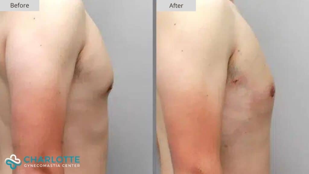
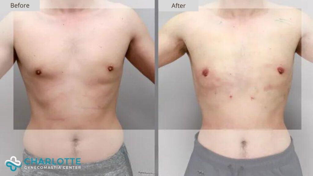
Instead of a flat and natural chest contour, he noticed hollowness beneath the nipples and an unnatural shape that became more apparent over time. From certain angles, the chest appeared indented rather than masculine. Despite being physically fit, the contour irregularity made his chest look incomplete and asymmetrical.
His concern was not that gynecomastia had returned. The issue was quite the opposite. Too much tissue had been removed during the original procedure.
Understanding Gynecomastia Over Resection
Over-resection is one of the more challenging complications of gynecomastia surgery. While excess glandular tissue must be removed to correct puffy nipples, preserving the correct amount of surrounding fat is just as important.
The male chest is shaped not only by skin and breast tissue, but by the pectoralis muscle anatomy underneath. In many men, the lower edge of the pectoralis muscle sits at or slightly above the visible chest crease. In others, the inferior border of the muscle lies higher, sometimes at nipple level when the muscle is contracted.
This patient had what is considered a high inferior border of the pectoralis muscle. In these cases, the fatty tissue beneath the nipple must be left slightly thicker to maintain a natural external contour. Removing both glandular tissue and too much fat in this area can lead to a sunken appearance.
That is precisely what occurred during his original surgery.
Why the Deformity Occurred
During the initial gynecomastia procedure, glandular tissue beneath the nipples was removed appropriately. However, fatty tissue under and below the nipple was also resected too aggressively. Since his pectoralis muscle did not extend far down the chest, there was no underlying structure to support the skin envelope.
As a result, the lower pole of the chest collapsed inward. The nipple appeared flattened and tethered. The natural curve between the upper chest and lower chest was lost.
This type of deformity cannot be corrected with additional tissue removal or skin tightening. The missing element is volume.
Surgical Planning for Correction
Correcting gynecomastia over-resection requires a restorative mindset. The goal is not to subtract, but to rebuild shape and balance.
For this patient, fat grafting was the ideal solution. Fat allows the surgeon to replace lost volume gradually and precisely, restoring a smooth contour without introducing foreign material.
The surgical plan focused on:
- Replacing volume beneath and below the nipple
- Softening the hollowed lower chest
- Recreating a natural transition from upper to lower chest
- Avoiding over correction while accounting for fat absorption
Since some fat is naturally reabsorbed by the body, the decision was made to intentionally overfill the area slightly. This ensures that once healing stabilizes, the remaining volume matches the desired contour.
The Fat Grafting Procedure
Fat was harvested from a donor area using gentle liposuction techniques. The fat was then carefully processed and prepared for reinjection.
Using small cannulas, fat was layered strategically beneath the skin in the deficient areas. Care was taken to distribute the fat evenly and avoid lumpiness. The focus was on restoring shape rather than creating projection.
This technique allows the chest to heal with a soft, natural feel that closely mimics native tissue.
Early Postoperative Appearance
The patient is shown one day after surgery, as he traveled from out of town and returned home shortly after the procedure. At this stage, swelling is expected and the chest appears fuller than the final result will be.
Even at this early point, the improvement in chest shape is visible. The lower pole no longer appears hollow. The nipple sits on a smoother surface rather than an indented one. The overall chest contour looks more balanced and proportionate.
As healing progresses and swelling subsides, some of the initial fullness will decrease. This gradual settling is anticipated and accounted for during surgical planning.
Why Revision Surgery Requires Experience
Revision gynecomastia surgery is fundamentally different from primary treatment. The anatomy has already been altered. Scar tissue may be present. Volume relationships have changed.
Successful correction depends on:
- Understanding male chest anatomy in detail
- Recognizing variations in pectoralis muscle positioning
- Knowing when to preserve tissue rather than remove it
- Using restorative techniques like fat grafting appropriately
This case illustrates how even well intentioned gynecomastia surgery can lead to deformity when anatomical nuance is overlooked.
Visual Outcome and Long Term Expectations
The early postoperative result shows a chest that looks fuller and more natural. Over the coming months, swelling will decrease and the grafted fat will stabilize. The goal is a chest that no longer draws attention to itself, allowing the patient to feel comfortable shirtless and in fitted clothing.
Since fat grafting uses the patient’s own tissue, the result integrates naturally with the surrounding chest. There is no implant and no rigid structure. The outcome evolves gradually rather than abruptly.
Restoring Balance After Over Correction
This case highlights an important truth about gynecomastia surgery. Removing tissue is only part of the equation. Preserving shape is equally critical.
For this patient, correction did not involve doing more of the same. It required reversing the imbalance created by over-resection and rebuilding what was lost. By restoring volume beneath the nipple and lower chest, the chest regains its natural contour and proportion.
This type of correction reflects a comprehensive understanding of male chest anatomy and the experience required to manage complications thoughtfully.